A breakthrough discovery in the treatment of glioma. All thanks to Polish scientists

Scientists from Bydgoszcz are working on more effective methods of diagnosing gliomas. Their discovery may revolutionize the treatment of patients and thus increase their chances of survival.
Gliomas are one of the most frequently detected tumors inside the skull. They arise as a result of abnormal growth of glial cells, which are responsible, among other things, for the protection and nutrition of neurons. So far, magnetic resonance imaging has been mainly used to diagnose this type of tumor. Unfortunately, this change detection method is not perfect. It does not allow for an accurate estimation of the size of the tumor, which makes subsequent treatment much more difficult and, as a result, reduces the patients’ chances of surviving and winning the fight against the disease. Fortunately, this may change soon. And all thanks to the efforts of Polish scientists from the Bydgoszcz University of Technology, headed by Professor Maciej Harat.
A new method of glioma imaging
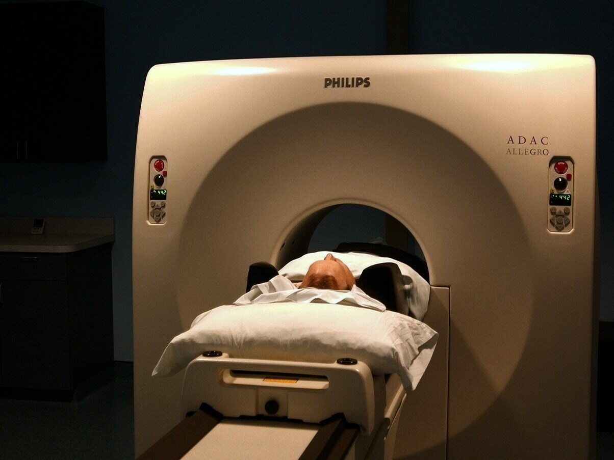
A team of experts from Bydgoszcz discovered that a more effective and more precise method of diagnosing glioma than magnetic resonance imaging is PET scanning, i.e. positron emission tomography (Positron Emission Tomography). This method is known and used in medicine. It involves administering harmless amounts of the rapidly decaying radioisotope fluorine 18F to the patient. After 20-40 minutes, tyrosine is introduced into the patient’s body. Molecules of this amino acid serve as a marker to mark the boundaries of the tumor. They are deposited in tumor cells, making it possible to locate them.
Experts from the Bydgoszcz University of Technology decided to slightly modify the PET examination procedure. The change involves administering tyrosine to the patient immediately after applying the radiotracer. The image of the glioma obtained by this method was much more accurate than those obtained using “traditional” PET or magnetic resonance imaging.
“During our work, we have shown that in each case we are able to determine the glioma infiltration beyond the area visible in magnetic resonance imaging. This indicates that even the most radical tumor resection using MRI imaging actually left a significant portion of the tumor behind, resulting in rapid disease recurrence. The discovery is a consequence of our main achievement, which was the verification of another brain imaging technique: PET examination using fluorine-labeled tyrosine,” emphasizes Professor Harat, the lead author of the study.
The effectiveness of the method proposed by scientists was confirmed by biopsies of brain tumors performed in patients with glioma at the Oncological Center at the 10th Military Clinical Hospital in Bydgoszcz.
Benefits of a new method of diagnosing glioma
A new method of diagnosing gliomas may increase the effectiveness of treatment for patients with brain tumors and reduce the risk of recurrence. The procedure for removing diseased tissues will become more precise, which will spare healthy cells.
“The collected evidence will allow us to better plan further clinical trials and hopefully, once introduced into clinical practice, we will finally achieve improved treatment results for these one of the worst-prognosis human cancers,” emphasizes Prof. Maciej Harat.


